Disease Agent of Nutty Fever: Bacillus Ugondie
In order to find the identity of our disease agent, a series tests were run. These tests determined whether our disease was a bacteria or virus, and the genus and species. The bacterial key used to find or disease agent's identity can be found at the bottom of the page.


|
Our genome size was determined by flow cytometry, which compared our disease agent's size to that of the K12 strain of E. coli. This strain has a genome size of 4.639221 Mb (Blattner et al. 1997).
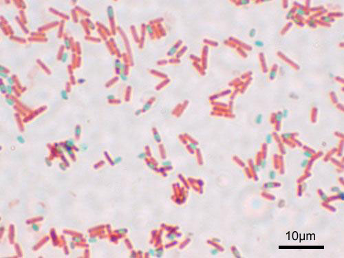
A virus would have a genome size less than 170 kb, while bacteria would have a genome size larger than 170 kb. We determined that our disease agent's genome size was about 4.068 Mb (4068 kb).This was much larger than a virus, making our disease agent a bacterium. Our first test was the Gram Stain and Shape test. A round shape indicates that the bacteria are cocci shaped, while a rod shape indicates a bacilli shape. A positive gram stain is shown by a blue/purple coloration, because of a layer of thick peptidoglycan on the outside of the bacteria. A negative gram stain will look pink, because the layer of peptidoglycan in thinner in these bacteria.
The results (purple coloration of our agent) indicated that it was gram positive and the shape was bacilli. The next test that was performed was the Spore test. This test detects the presence of endospore (spores that form within the bacteria. A positive result will show a bluish-green area within the bacteria, while a negative result will show nothing.
The results of our tests were positive, as can be seen in the picture. The Aerobe/Anaerobe test was performed on our disease agent. A white coloration in the test tube indicates that the bacteria are growing in that area. Growth in the top but not the bottom indicates that the bacteria is an obligate aerobe. Growth in the bottom but not the top indicates that it is an obligate anaerobe. Uneven growth throughout indicates that it is a faculative aerobe.
Our disease agent was shown to be a faculative aerobe. According to our bacteria was in the genus Bacillus. The Starch Hydrolysis test was performed on our disease agent. This test was to detect whether the bacteria can use its enzymes to hydrolyze and use starch. Iodine is put into the starch agar to give it a purple color that can detect starch hydrolysis. A negative result will show no change in the purple coloration, but a positive result will result in a lightening of the color around where the bacteria was placed.
Our disease agent shows a yellow coloration where the bacteria was place, indicating a positive result, as can be seen in the picture. |

|
The final test our group performed was the Voges-Proskaur test. This test determines whether the disease agent produced butandiol (a neutral end product) during glucose metabolism. A positive result will evolve a pink color while the negative result will remain relatively colorless.
Our disease agent gave a positive result, keying out to be an unknown Bacillus species. This newly discovered species was then named Bacillus ugondie, which causes Nutty Fever. |
| bacterial_key.pdf |
Nutty Fever: Disease Classification
The disease type for Nutty Fever is bacterial meningitis that is caused by our bacterium, Bacillus ugondie.
General bacterial meningeal symptoms such as fever, headache, and changes in mental status, are a common occurrence among patients with Nutty Fever. More specific meningeal like nuchal rigidity, cranial nerve palsy, Kernig's sign, and Brudzinski's sign are also characteristic of the disease.
The lack of voluntary motor control found in bacterial meningitis patients was also seen in Nutty Fever patients. This is exemplified by the ataxia and nystagmus developed by patients with Nutty Fever. As Nutty Fever progresses, the advanced symptoms (bradycardia, coma, and palsy of cranial nerve III) are the same as those commonly found in cases of bacterial meningitis as well (Schlossberg, 2008).
A rash on the extremities can also be found in cases of bacterial meningitis, as was found in the petechial rash of the arms and legs of several patients with Nutty Fever.
References:
Blattner F.R. et al. 1997.The complete genome sequence of Escherichia coli K-12. Science. 227(5331): 1453-1462.
Schlossberg D. 2008. Bacterial meningitis, pp. 505-512. Clinical infectious disease. Cambridge University Press, New York, NY.
Blattner F.R. et al. 1997.The complete genome sequence of Escherichia coli K-12. Science. 227(5331): 1453-1462.
Schlossberg D. 2008. Bacterial meningitis, pp. 505-512. Clinical infectious disease. Cambridge University Press, New York, NY.
